Sternal fractures – Have you ever been in a motor vehicle accident (MVA), had a pedestrian accident, had CPR performed on you, speared in the chest while playing football? Did you have intense chest pain? Did you have difficulty breathing afterwards? If you said yes, then there was a strong possibility that you fractured your sternum.
Causes
● Sternal fractures are associated with blunt force trauma to the chest.
o Before the passage of the seatbelt laws, sternal fractures occurred in approximately 3% of motor vehicle accidents (MVAs). Of course, the percentage has declined over time.
● Sternal fractures can be caused by direct impact sports, falls, being hit by a car, assault.
spontaneous stress fractures, and osteoporosis and kyphosis of the thoracic spine, especially in women.
● Long-term steroid use can also cause a sternal fracture.
● CPR may cause a sternal fracture.
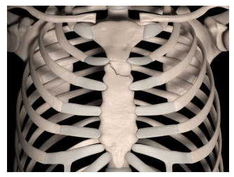
Sternal fractures appear in the midbody and transverse area of the sternum (breastbone). Manubrium fractures are the most common. Injuries have a mortality rate of less than 1% because of chest injuries like cardiac contusions, aortic rupture, pulmonary contusion, and thoracic spine compression fractures.
Symptoms
● A sternal fracture can cause localized pain or pain over the entire chest.
o If someone is experiencing this type of pain, it is extremely important to call 911 and go directly to the nearest ER to rule out myocardial infarction (heart attack).
● Difficulty breathing is present in 15% to 20% of the patients, which may be caused by cardiopulmonary contusion.
Diagnosis
● Once in the ER, a cardiac work-up to rule out a heart attack will begin.
● EKGs will be repeated every 6 hours.
o However, if the EKG results are normal, no further work-up is required.
● Anterior, posterior, and lateral view X-rays will be performed if there is suspicion of sternal fracture.
o Anterior- posterior view X-ray can be helpful in detecting other injuries: rib fractures, pulmonary contusion, hemothorax, and pneumothorax.
● A chest CT is the most common image study to assess the degree of sternal displacement as well as other chest injuries.
Treatment
● Surgery fixation is not necessary for patients with sternal fractures.
● Treatment can be performed outpatient unless other injuries are associated with the injury.
● Pain management follow-up may be necessary for medication and NSAIDs.
● Patients tend to recover quickly (several weeks’ time) from isolated sternal fractures.
● Sternal fractures in children should raise suspicion for abuse.
● Please follow-up with a pediatrician, family physician, or nearest ER as soon as possible.
● If you suspect that the child may be in danger, please call the police and child protective services.
References
Felten, S. (2017). Sternal Fracture Differential Diagnoses. https://emedicine.medscape.com/article/826169-differential
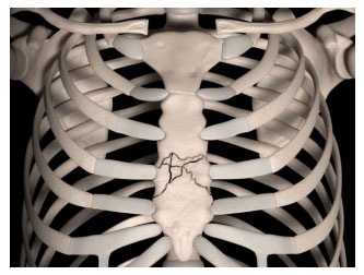
Marchal, D. (2020). [Illustration]. Digital medical illustration depicting a comminuted fracture of the breastbone (sternum). Anterior (front) view. 3D rendering. https://www.shutterstock.com/image-illustration/digital-medical-illustration-depicting-comminuted-fracture-1399874501
Marchal, D. (2020). [Illustration]. Digital medical illustration depicting a fracture of the breastbone
(sternum). Anterior (front) view. 3D rendering. https://www.shutterstock.com/image-illustration/digital-medical-illustration-depicting-fracture-breast-1399874504
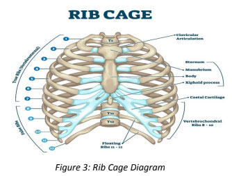
VectorMine. (2020). [Illustration Diagram]. Rib cage anatomy, labeled vector illustration diagram. Medical human chest skeletal bone structure model. Numbered ribs, sternum, cartilage parts and clavicular articulation. Health care education. https://www.shutterstock.com/image-vector/rib-cage-anatomy-labeled-vector-illustration-1650017491