A flail chest is a medical emergency and results from severe blunt trauma to the ribs and chest in multiple areas. The trauma can be caused by a MVA (rollover injury or crash), falls, and assaults in younger, healthier patients. Lesser trauma includes osteoporosis, total sternotomy, multiplemyeloma, and congenital absence of the sternum. Elderly patients are predisposed because of age-related physiological stiffness of the chest wall and because they may have osteoporosis. They may also have a per-existing lung disease that may make them a high risk for complications. It is the abnormal inward movement of a section of the chest wall caused either by more than 3 rib fractures anteriorly or posteriorly within each rib. A flail chest is clinically diagnosed when a portion of the chest wall moves separately from the rest of the chest wall while inhaling and exhaling and often unilateral but can be bilateral and there are multiple variations like posterior flail segments, anterior flail segments, and flail sternum with ribs on both sides of the thoracic cage fractured, which may cause heart and lung damage. A decline in respirations because of a flail chest is likely caused by severe pulmonary contusions (bruised lungs) that damage the chest wall.
There is a 5% to 10% mortality rate if patients reach the hospital alive. Flail chest injuries are often accompanied by pulmonary contusions, hemopneumothorax, heart injury, and an occasional major vascular injury. Patients who do not need mechanical ventilation do better. Mortality increases with increased injury severity, age, and total number of rib fractures. Patients who are suspected of having a flail chest will be worked-up in the ER by having X-rays with rib series and a sagittal and coronal view of the thoracic spine by a multi-slice computed tomography (MSCT) scan of the chest to assess for rib fractures. Blood work will include Arterial Blood Gases (ABGs) to assess the severity of hypoventilation caused by a possible pulmonary contusion (lung bruise) and the pain caused by the rib fractures. The results of the work-up will help determine the need for mechanical ventilation. Mechanical ventilation is used for patients who have persistent respiratory insufficiency, failure after adequate pain control, or complications related to excessive narcotic administration. Medical treatment will require adequate pain control and management of the pulmonary injury prior to being placed on mechanical ventilation.
Surgical treatment typically requires an operative fixation. An operative fixation is determined by the severity of the underlying pulmonary contusion versus chest wall instability, multiple myeloma, sternal absence, or total sternotomy.
A flail chest diagnosed in children is a potential sign of child abuse. Please follow-up with a pediatrician, family physician, or nearest ER as soon as possible. If you suspect that the child may be in danger, please all the police and child protective services.
Bjerke, H.S. (2016). Read more
Richelle, Y. (2020). Flail chest: do you know about it? yes or no. [Photo]. Read more
Solar22. (2020). Anatomy of human rib. Illustration from vector about science and medical. [Diagram]. Retrieved from https://www.shutterstock.com/image-vector/anatomy-human-rib-illustration-vector-about-600282818.
Udekwu P, Roy S, McIntyre S, Farrell M. Influence on Length of Stay and Mortality in BluntChest Injury. Am Surg. 2018 Sep 01;84(9):1406-1409.
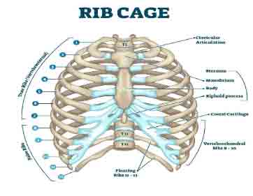
VectorMine. (2020). [Illustration Diagram]. Rib cage anatomy, labeled vector illustration diagram.Medical human chest skeletal bone structure model. Numbered ribs, sternum, cartilage parts andclavicular articulation. Health care education. Retrieved from https://www.shutterstock.com/image-vector/rib-cage-anatomy-labeled-vector-illustration-1650017491