Experiencing neck pain? Cervical (neck) pain has many causes. Whiplash related to a motor vehicle accident, pulling on your neck while doing sit-ups, using the arm of your couch as a pillow while lying down, habitual neck popping, wrestling, playing football or soccer, and jamming your neck into a pillow while sleeping are just a few common examples. Chronic neck stiffness may be caused by cervical lordosis, which is the straightening of the cervical spine and/or scoliosis, the curving of the spine. These conditions can lead to stiffness and occasional or constant headaches. Neck pain can also be idiopathic which means there is an unknown cause for the pain. One morning you wake up and notice that it is difficult to look over your shoulder; you have a mild headache that doesn’t go away, or you have numbness, tingling, dull, achy, electrifying, sharp, or stabbing pain that radiates into either shoulder, down your arm, and into your fingers.
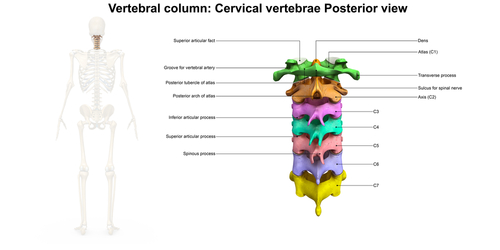
In this article, I will describe the most common types of neck pain that I see as a pain management nurse practitioner and explain how I treat it at my clinic. Before I begin, it’s important to discuss basic cervical anatomy (Figure 1). The neck begins at the base of the skull. It consists of seven vertebrae (bony structures) and five discs, which act as shock absorbers and prevent the vertebrae from rubbing together. The brainstem (spinal cord) runs down the length of the spine ending in the lumbar (lower back) area. The nerves branch off the spinal cord and pass through the foramina to innervate the muscles.
The best way to determine the origin of cervical pain is a magnetic resonance imaging (MRI) or computed tomography (CT). MRIs are more detailed than CT scans. However, both scans can show the tissue around the specified image, bone structure, and nerves. If you visit your primary care provider for neck pain, you will receive an X-ray first because insurance companies require it. If the cervical pain continues to persist, an MRI needs to be ordered. A CT can be ordered if patients have a metal implant (stents, pacemakers, and intrathecal pain pump).
The X-ray, MRI, and CT scans report identify areas of the spine in sections. Since I’m discussing the cervical spine, let’s talk about dermatomes. In short, dermatomes are areas of the skin connected to the spinal nerve. Dermatomes pinpoint exactly where pain originates in the body.

C2 (The main function of this section is to allow the neck to rotate. Trauma to this area can cause chronic headaches, muscle stiffness, and difficulty turning your head from left to right.)
C2-C3 and C3-C4 (These areas are in the stem of the neck. Centralized neck pain and stiffness may be felt in this area.)
C4-C5 (Located at the base of the neck where the neck meets the shoulders, C4 supplies nerves to the shoulders and C5 supplies nerves to the deltoids.)
C5-C6 (C5 supplies nerves to the deltoids and C6 nerves stimulate the biceps.)
C6-C7 (C6 nerves stimulate the biceps and C7 nerves stimulate the fingers.)
Neuropathic pain (nerve pain) can radiate down the neck, into the shoulders, biceps, and the fingertips depending on the location of bulging discs pressing on the nerves. I will discuss neuropathic pain in detail in future articles. Also, in addition to using dermatomes to explain where pain originates in the neck, dermatome maps can be used to pinpoint pain located anywhere in the body. Please google dermatome maps, select “images,” and scroll down until you see dermatomes with a color-coded key. This type of dermatome map will come in handy when you try to read either your CT or MRI on your own, as you try to pinpoint the source of your pain.
References:
Kayalioglu ,Gulgun. (2009). The Spinal Cord. Retrieved from https://www.sciencedirect.com/topics/medicine-and-dentistry/dermatome
Sciencepics. (2019). Cervical spine posterior view 3d illustration. [Illustration].
Stihii. (2019). Medical, didactic board of anatomy of human sensory innervation system, dermatomes and cutaneous nerve territories, segmental, radicular, cutaneous innervation of the anterior trunk wall. [Drawing]
